

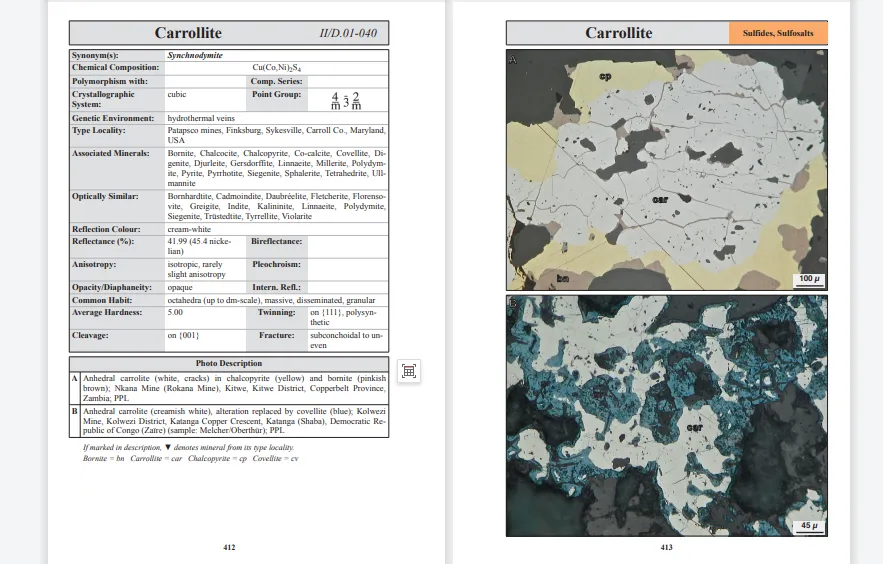
-
About Us
-
-
-
Contact Us

Microscopic images of metallic minerals—Heijinggang Reports
Reflection Light Polarizing Microscope Identification
The reflected-light polarizing microscope is the primary tool for identifying opaque metallic minerals. By examining characteristics such as the mineral's reflection color, reflectivity, pleochroism, internal reflections, and homogeneity—along with its physical properties and associated mineral assemblages—minerals can be accurately identified.
Reflective color and reflectance Different metal minerals exhibit distinct reflection colors and reflectance levels. For instance, chalcopyrite reflects a yellowish-white hue, while tetrahedrite displays a grayish-white color with subtle creamy-yellow tints.
Multicolourness and Internal Reflection Some minerals exhibit pleochroism or internal reflection characteristics under reflected light, features that can be used to distinguish between minerals.
Homogeneity and Heterogeneity : Observe the homogeneity of minerals under reflected light—non-homogeneous minerals may exhibit distinct internal reflections.
Applications of the Microphotometer
A microspectrophotometer can be used to measure the reflectance of minerals, further enhancing the accuracy of identification. By determining the reflectance values of minerals and combining them with other optical characteristics, it becomes possible to narrow down the range of mineral species.
Optical Properties Under a Polarizing Microscope
For transparent or partially translucent minerals, a polarizing microscope can be used to observe their optical properties, such as interference colors, birefringence, pleochroism, and extinction types.
Interference Color and Birefringence By observing the interference colors and birefringence of a mineral, one can determine its axial character and optical sign.
Ductility and Extinction Types : Ductility refers to the variation in interference colors of a mineral under polarized light, while the extinction type reflects the mineral's optical properties.
Hyperspectral Imaging Technology
Hyperspectral imaging technology combined with microscopy can be used for the automated identification of metallic minerals. By capturing images of mineral samples with a hyperspectral camera and analyzing their spectral characteristics, rapid identification is achieved.
Other auxiliary methods
Erosion reaction : Certain minerals undergo etching reactions when exposed to specific chemical reagents, and observing the reaction characteristics can help with identification.
Mineral morphology and coexisting relationships Observing the mineral's morphology—such as euhedral, subhedral, and other crystal forms—as well as its symbiotic relationships with other minerals, also serves as an important basis for identification.
Summary
Identifying metallic minerals under a microscope requires the integrated application of multiple methods and techniques, including reflected-light polarized microscopy, microspectrophotometry, polarized light microscopy, and hyperspectral imaging. By examining the minerals' optical properties, physical characteristics, and structural features—along with their associated mineralogical relationships and chemical reactions—it becomes possible to accurately identify metallic minerals.
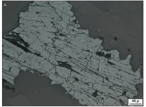
Molybdenite from the Bogala Mine in Sri Lanka
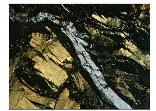
Skeleton copper (a soft, delicate light pink), paired with a touch of copper ore (a subtle bluish-gray).
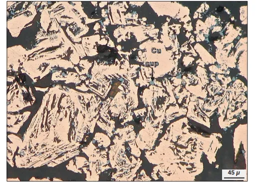
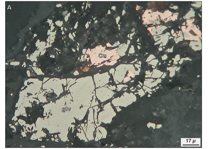
Amorphous native copper (darker in color, with visible scratches) fills fine veins within jadeite (which exhibits red reflective light internally); from the Black Sea region of Turkey, Rize Province, Çayeli District, Madenköy Mine (sample: Harbach); observed under a polarizing microscope.
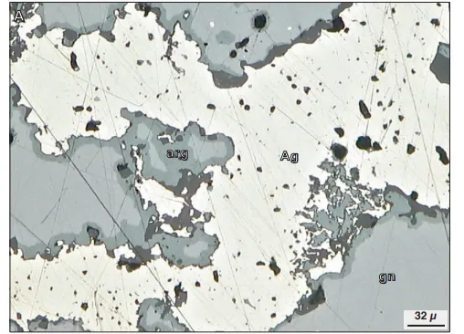
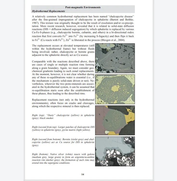
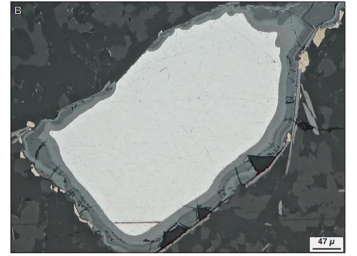
Natural silver (white) without visible crystals is embedded within galena (light gray; mineral abbreviation "gn"), featuring a thin-layered argentite reaction rim (gray-green; mineral abbreviation "arg").
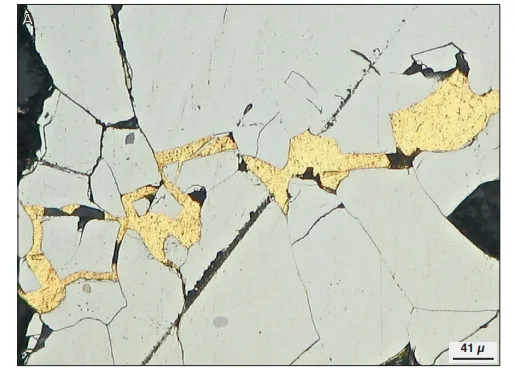
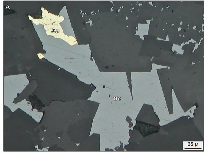
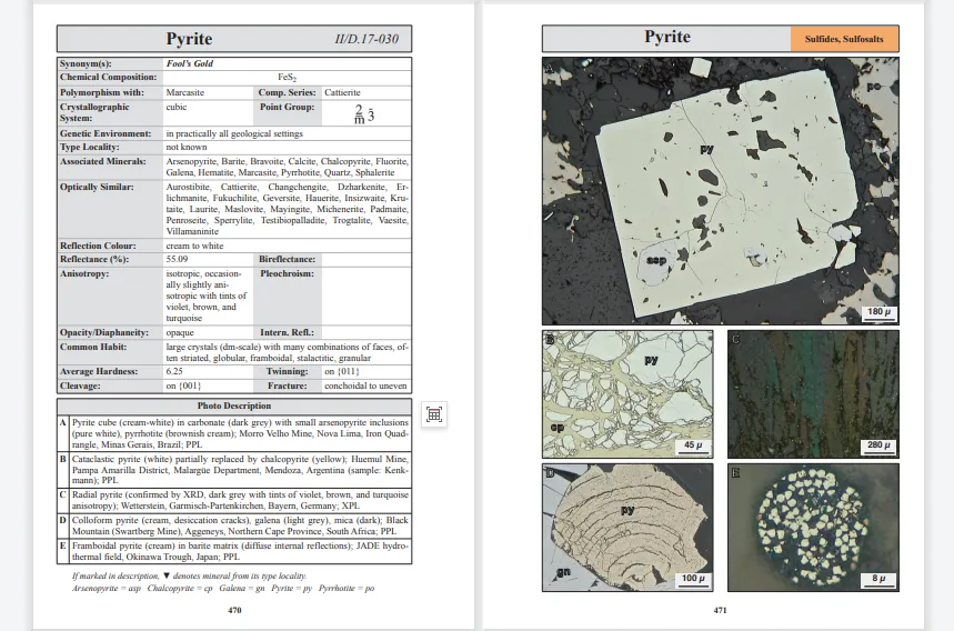
Native gold (yellow) fills the fractures in pyrite (light cream-colored); where the two minerals meet, the pyrite appears particularly dull. Gauteng Province, South Africa, Witwatersrand region
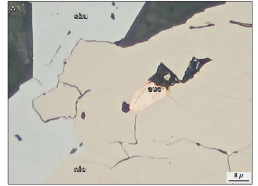
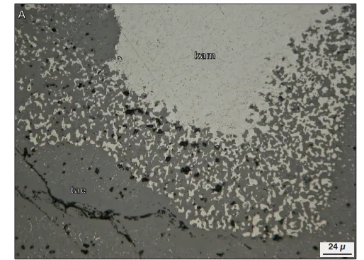
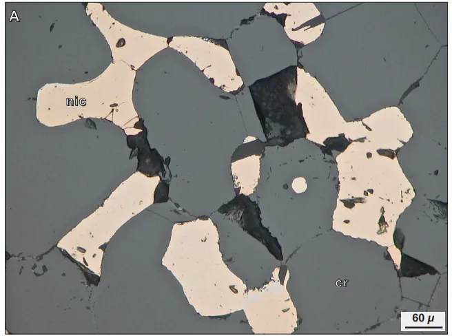
The subhedral chalcopyrite (bright orange) is embedded within a nickel ore matrix (slightly pinkish-brown, with weak anisotropy), accompanied by niccolite (light gray).
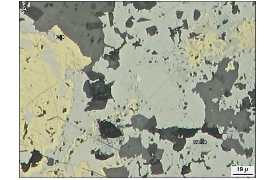
The cinnabar-argentite ore (beige) occurs intimately associated with chalcopyrite (yellow), surrounded by dark gray gangue minerals; Landsburg, Arzfeld-Obermoschel, Rhineland-Palatinate, Germany
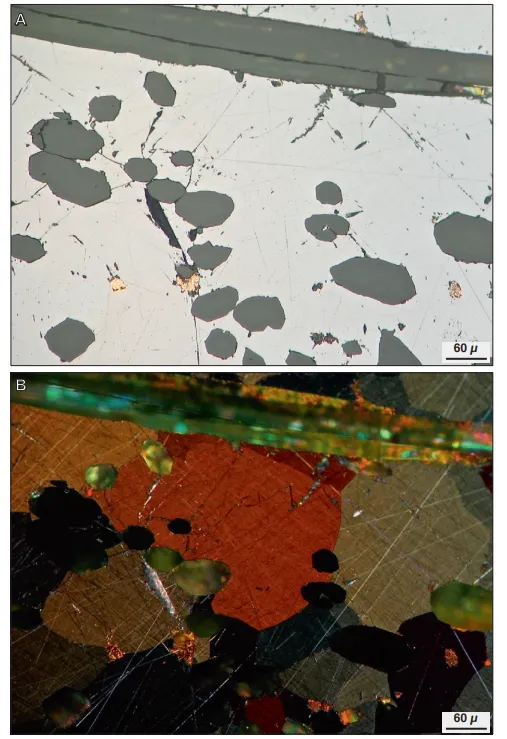
Natural lead without visible crystals (medium gray with brownish hues, exhibiting oriented lamellae) occurs in association with dolomite (dark gray); Hällstigen Mine, Päseberg area, Filipstad Municipality, Värmland County, Sweden
A network-like structure composed of native iron (light gray) is embedded within a silicate matrix (dark gray); the sample was collected from Kekertarsuaq Island in Qaasuitsup Municipality (West Greenland), located off the western coast of Greenland.
Iron without self-formed crystals (light gray), featuring goethite rims (in various shades of dark gray), with ilmenite (brown) distributed along the edges of iron oxide particles, and silicate minerals (dark gray).
Sphalerite (mottled light gray) with euhedral crystals and dense, elongated granites (dark gray); meteorite
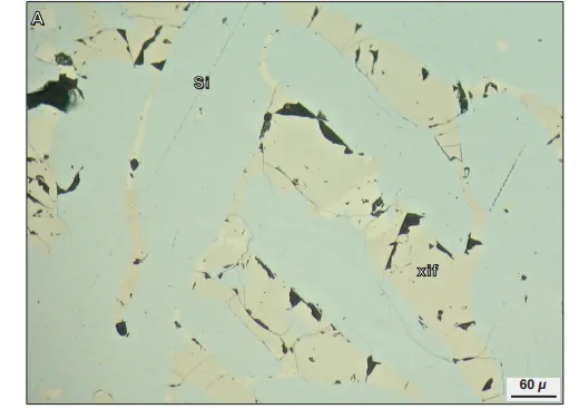
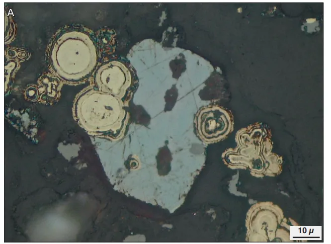
Xifeng mineral (light brown) is embedded within a silica matrix (with subtle gray tones); inside the Xifeng mineral at the center of the field of view, there exists another unidentified mineral (slightly lighter in creamy hue).
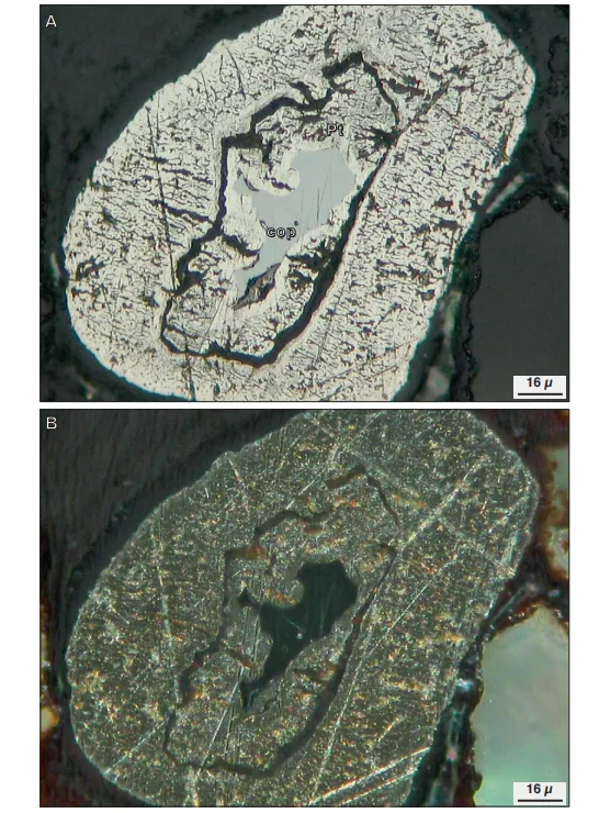
Euhedral iridium inclusions (white) within heteroplatinite (slightly tinged with white), partially bordering the lower sections of chalcopyrite (medium gray).
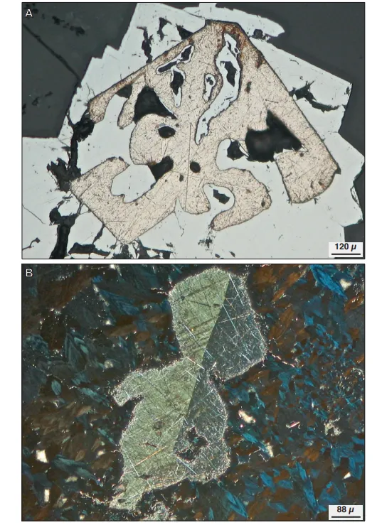
Porous particles of native platinum (milky white) forming the edges around a copper ore deposit (light gray).
Naturally occurring bismuth, featuring an euhedral contact twin structure (with distinct anisotropy along the twin boundaries), also reveals several faint diagonal twin lamellae in the brighter upper-left region. The matrix is composed of tetrahedrite (showing brown-to-blue anisotropy).
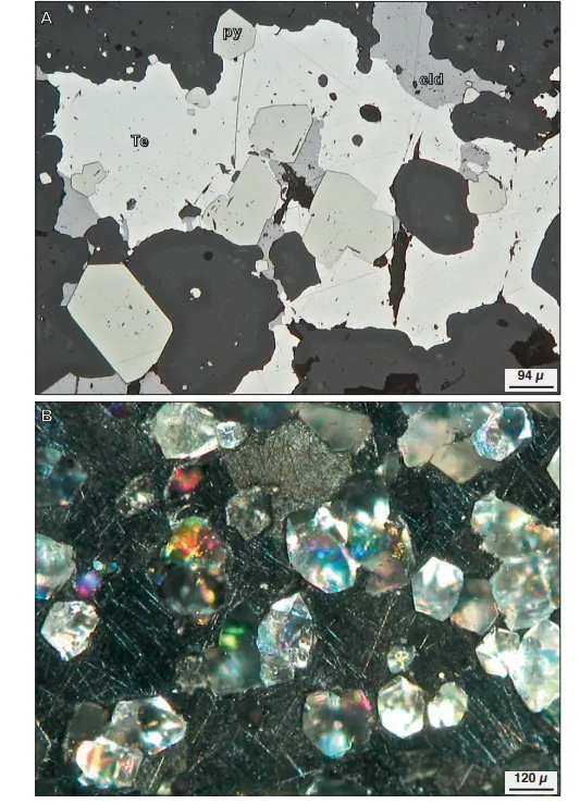
A massive natural tellurium (anisotropic, displaying various shades of brown and gray) forms the matrix, within which are embedded well-formed quartz crystals exhibiting bright internal reflections.
Copper-antimony ore (with gray, brown, and red internal reflections), native copper (with faint orange reflections), mica, and other euhedral silicate minerals (showing multicolored internal reflections).
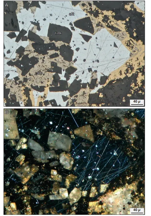
Tischendorfite mineral, with large vesuvian crystals (light gray) at the edges, along with euhedral to subhedral ferroan dolomite (dark gray) and subhedral hornblende (light brown).
Aphyric chalcocite (blue-gray) replaces pyrite (light yellow), with both minerals embedded within a gel-like goethite (in varying shades of dark gray); minor associated azurite (blue) is also present.
The colorless godlevite (a slightly olive-hued gray—no pleochroism observed here, with irregular fractures)—is closely associated with native copper (pinkish) and silicate minerals (ranging from dark gray tones).
The selenium mercury mineral (blue-gray), which lacks a distinct crystal form, occurs as an inclusion within a carbonate matrix (dark gray, with faint bi-reflection), and is found in association with native gold (bright yellow).
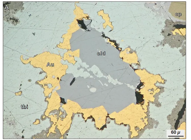
Tellurium mercury ore (medium gray), gold (yellow), bismuth telluride ore (light gray-green), and chalcopyrite (olive green, located in the upper right corner) occur together without self-formed crystals.
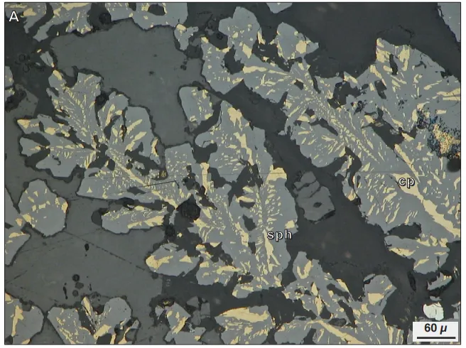
Epigenetic skeletal intergrowths of chalcopyrite (yellow) with sphalerite (medium gray) and anhydrite (medium gray, but darker than sphalerite); JADE hydrothermal field in the Okinawa Trough, Japan
Layered exsolution structures of chalcocite (gray) and cuprogallite (grayish purple) are present within chalcopyrite (yellow).
Massive tin-lead ore (olive-gray, with typical cleavage), pyrite (cream-colored), and tetrahedrite-sphalerite-argentopyrite (light gray) occur together; Ito Mine, Serado Province, Oruro City, Oruro Department, Bolivia 
Massive cinnabar (medium gray-green), galena (light gray), pyrite (creamy white), chalcopyrite (dark yellow), and quartz (dark gray, euhedral crystals) occur together.
Automorphic argentopyrite (bluish-gray) occurs in association with sphalerite (dark gray) and chalcopyrite (deep yellow), along with well-formed quartz crystals (dark gray); Mouzaía area, Tipasa Province, Algeria
Cinnabar crystals (bluish-gray) occur in association with gel-like pyrite (creamy white), embedded within an amorphous silica matrix (dark gray).
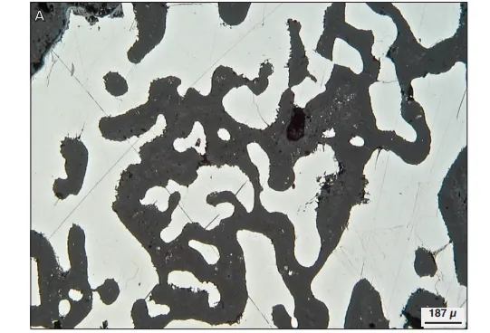
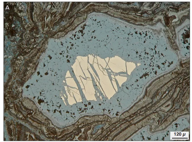
Nickel ore (pink) in the interstitial spaces between chromite grains (gray); Jezzine peridotite body in Ojén, Málaga Province, Andalusia, Spain
The material is sourced from the internet.
Previous Page
